Kingdom Monera - Structure of Bacterial Cell

Description:
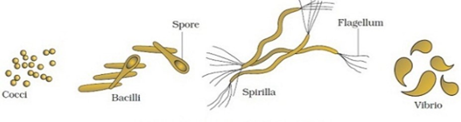
Average size of a typical bacterial cell ranges from 2.0 - 2.6 micrometer in length and 1.1 - 1.5 micrometer in width. On the basis of shape bacterial cells can be of the following types −
Coccus − Spherical or ovoid in shape. They can be monococcus (occurring singly), diplococcus (in twos), tertacoccus (in tetrads), streptococcus (in chains), staphylococcus (irregular grape like clusters) and sarcina (3-dimentional geometric forms).
Bacillus − The bacterium is straight and cylindrical like a rod with ends being flat, rounded or cigar shaped.
They can be diplobacillus (in twos) or streptobacillus (in chains).
Spirillum − Coiled like a cork-screw.
Vibrio − Comma or curved rod shaped. e.g. Vibrio cholera.
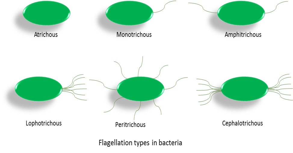
Depending on the presence or absence of flagella, bacteria can be grouped into flagellated and non-flagellated types.
The various forms of flagellation can be of the following types −
Atrichous − Flagella absent.
Monotrichous − A single flagellum occurs at or near one end of bacterium.
Amphitrichous − A flagellum at each of the two ends.
Lophotrichous − A group or tuft of flagella is found only at one end.
Cephalotrichous − A group or tuft of flagella at each of the two ends.
Peritrichous − A number of flagella are distributed all over the surface.
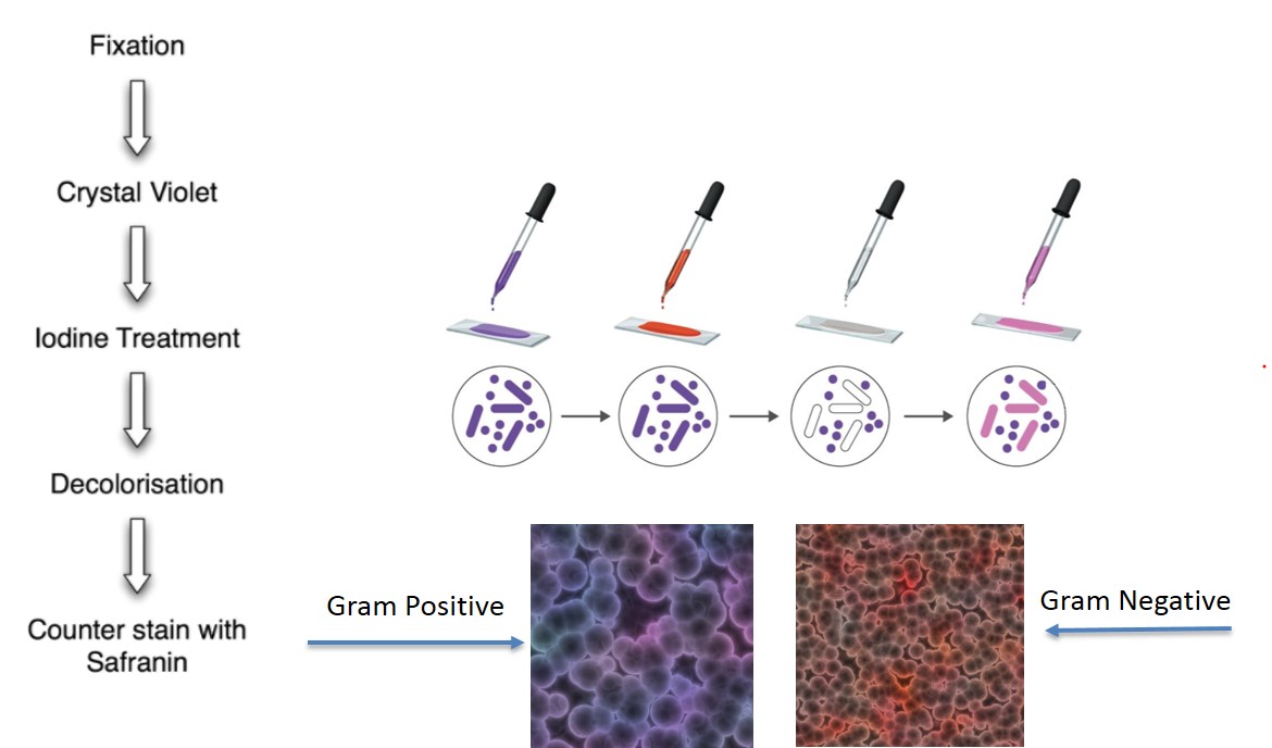
Bacteria has been classified into gram positive and gram negative bacteria based on the reaction of bacteria to Gram staining.
Procedure of Gram Staining −
Cells are first stained with an alkaline solution of crystal violet.
After sometime the cells are treated with 0.5% iodine solution followed by washing with water and alcohol or acetone.
Cells that take up the colour of crystal violet appears blue or purple under microscope and are known as gram positive bacteria.
The cells are then treated with a counter stain called safranin which is red in colour. The cells that did not take up the crystal violet stain, they are stained red or pink and are called gram negative cells.
Mechanism of staining −
| Gram positive bacteria | Gram negative bacteria |
|---|---|
| Thick peptidoglycan layer | Thin peptidoglycan layer |
| Crystal violet form a complex with iodine, CV-I complex, and this complex gets intercalated in between the thick peptidoglycan layer. | CV-I complex is formed but cells mostly cannot retained it because of thin peptidoglycan layer. |
| Washing with alcohol dehydrates the cells and the peptidoglycan layer shrinks hence the CV-I complex gets tightly stuck in between them. | Alcohol treatment removes the stain. |
| Safranin stain is not taken up. | Safranin stain is taken up by these cells and appear red under microscope. |